
FMGE Pathology Image Based Questions
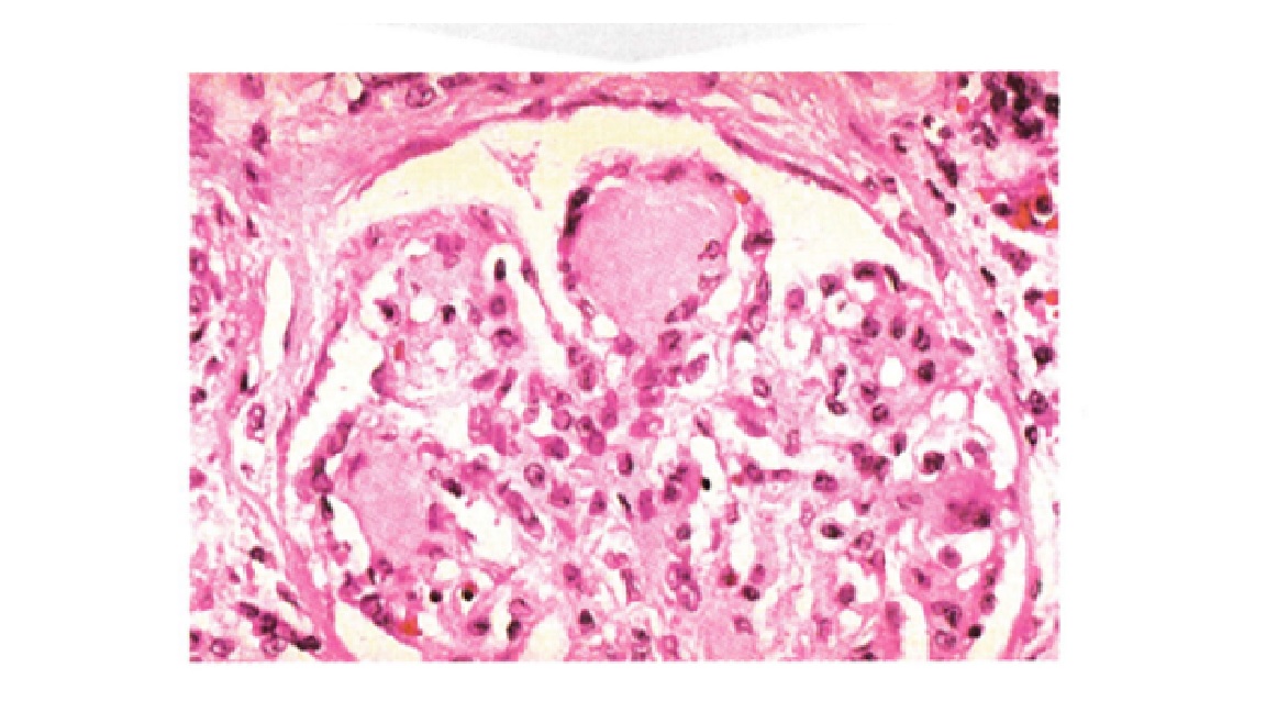
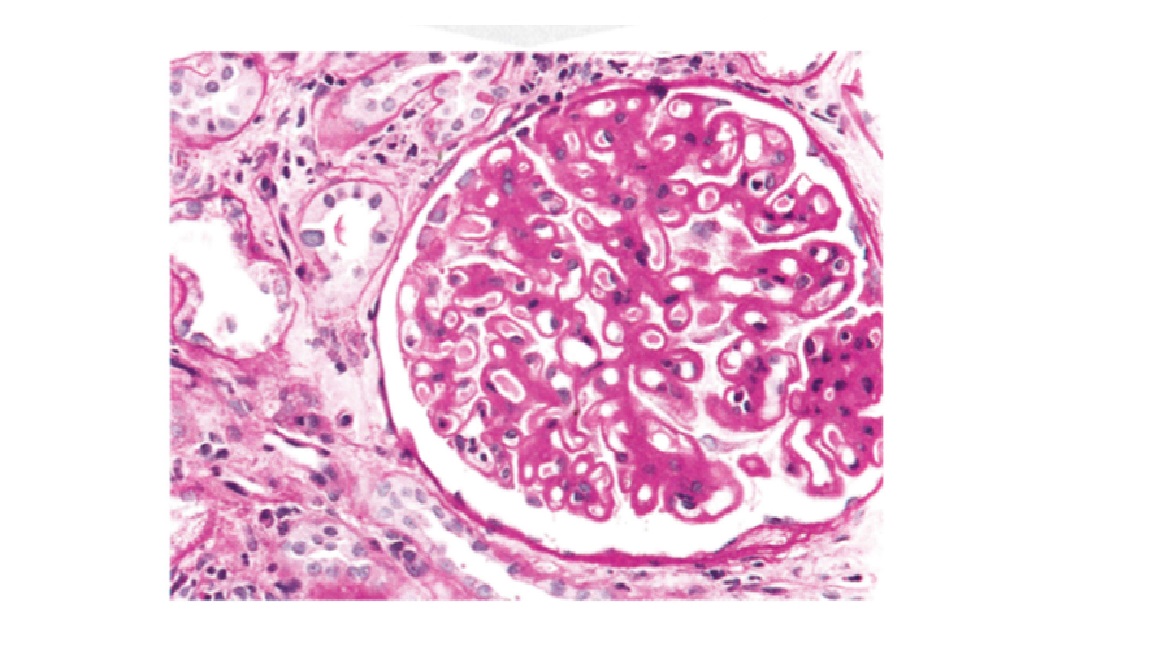
In Emergency department, a 50-year-old male presented with blurring of vision. Proteinuria is found on urine examination. Upon fundus examination there were dot and blot haemorrhages, microaneurysm and cotton wool spots seen. Histopathology picture of kidney given below. Make a diagnosis
Correct!
Wrong!
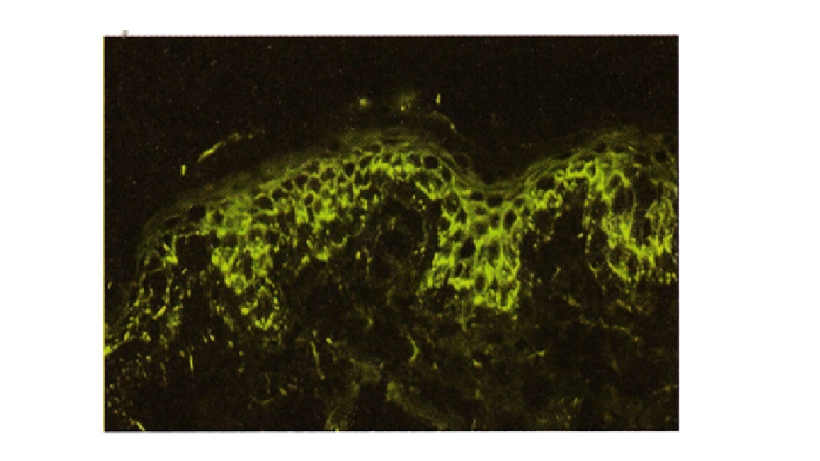
In OPD, a 26-year-old woman presented with painful blister in skin and oral mucosa. Direct immunofluorescence photograph is provide below. False statement is
Correct!
Wrong!
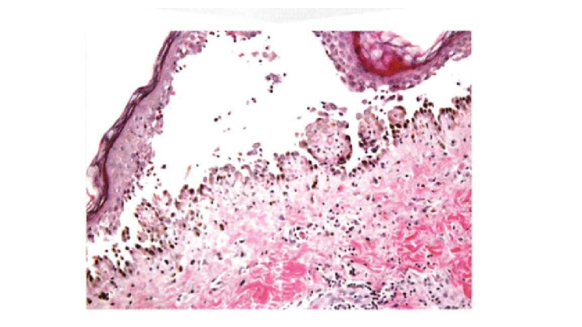
Diagnosis of the histopathology Photograph
Correct!
Wrong!
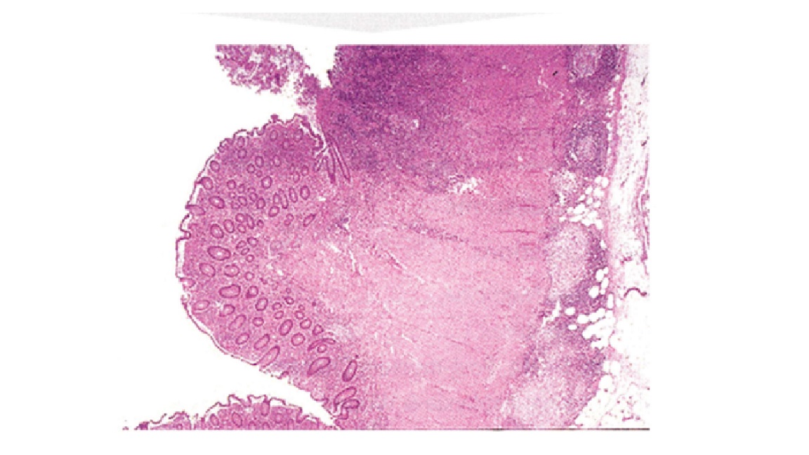
In Emergency department, a 50-year-old male presented with recurrent bloody diarrhea. Patient's colonoscopy test showed geographical ulcers. Histopathology is given in Photograph below. Your diagnosis is
Correct!
Wrong!
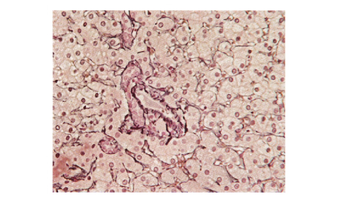
Staining is done for a Section of Liver (Photograph). Identify the stain
Correct!
Wrong!
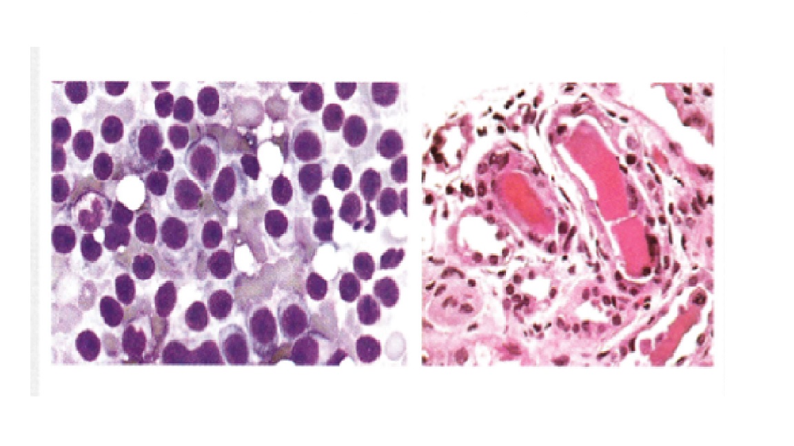
In Emergency department, an elderly male presented with history of intractable diarrhea. Patient' bone marrow and renal biopsy Photograph is given below. Most appropriate diagnosis will be
Correct!
Wrong!
In OPD, a 56-year-old male with frothy urine and facial puffiness. DIF and electron microscopic Photograph is given. Your diagnosis
Correct!
Wrong!
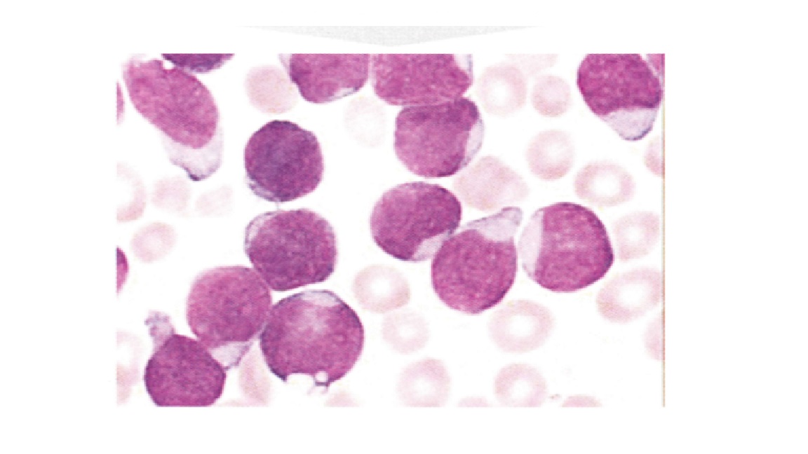
Eight years old child presents with fever, weight loss. On examination he was found to be pale with significant lymphadenopathy. Bone marrow histology is shown below in Photograph. Make a diagnosis
Correct!
Wrong!
38 years old male presented to OPD with fever with enlarged, ulcerated tonsils. PBS showed lymphocytosis with Monospot test was negative. Tonsillectomy carried out and it showed large cells mixed with lymphocytes. Cells were shown to be positive for CD20, EBVLMP1, MUM1, CD 79a, and background cells were positive for CD3. The cells were also negative for CD 15. Most likely diagnosis
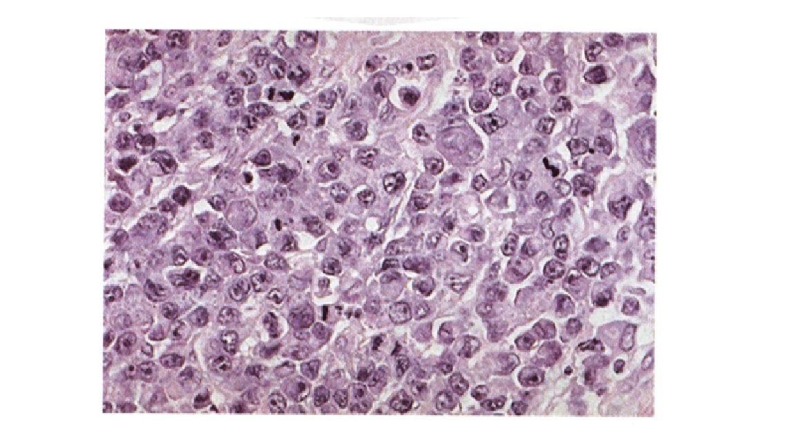
Correct!
Wrong!
45years old male patient presented to Emergency wing with heart burn and increased salivation. Upper GI Endoscopy carried out, biopsy was taken (See Photograph below). Most likely diagnosis is
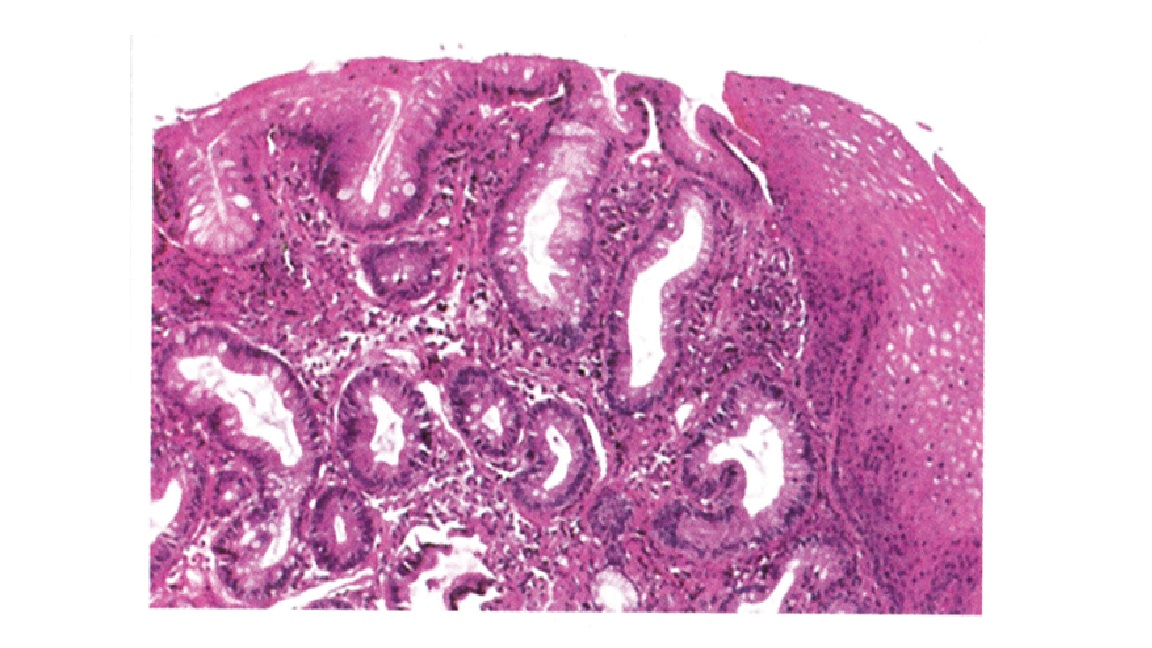
Correct!
Wrong!
FMGE Pathology Image Based Questions


| How to Prepare Pathology for FMGE? | How to Prepare Pathology for FMGE? |
 |
 |
Related Tests: OBG FMGE Past Paper | Anesthesia FMGE Past Paper | Orthopedic FMGE Past Paper